Back Of Neck Anatomy Muscles : The Intrinsic Back Muscles - Attachments - Actions ... - They start at the top of the neck and go down to the tailbone.. The posterior muscles of the neck are primarily concerned with head movements, like extension. The suprahyoid muscles originate from above the hyoid bone in the chin region. Rectus capitis, longus capitis, longus colli. Alle muscles are detailed described incl. Almost every muscle constitutes one part of a pair of identical bilateral.
The head rests on the top part of the vertebral column, with the skull joining at c1. Anterior muscles of the neck. Beneath the integument the back of neck presents in the median plane the ligamentum nuchae, which is a triangular fibrous sheet and represents upward the muscles of entire back are arranged in three groups—superficial, intermediate and deep (fig. They are divided into three groups, as shown below. Muscles of the shoulder and back laminated anatomy chart.
Learn Muscle Anatomy: Scalene Muscles and Other Neck Anatomy from www.visiblebody.com Human muscle system, the muscles of the human body that work the skeletal system, that are under voluntary control, and that the following sections provide a basic framework for the understanding of gross human muscular anatomy, with descriptions of the large muscle groups and their actions. The muscles of the shoulder and back chart shows how the many layers of muscle in the shoulder and back are intertwined with the other relevant systems and muscles in adjacent areas like the spine and neck. Spinous processes of txi to liii and supraspinous ligaments. Anterior muscles of the neck. Last update october 2, 2020. The three scalene muscles are found forming the floor of the posterior triangle. The muscles of the back that work together to support the spine, help keep the body upright and allow twist and bend in many directions. The anterior muscles of the neck facilitate swallowing and speech.
There are many muscles around the neck that help to support the cervical spine and allow you to move your head in different directions.
They move the head in every direction, pulling the skull and jaw towards the shoulders, spine, and scapula. Bones of the neck picture. As you know, the neck is the part of the body that sits between the head and torso. The neck muscles (and neck anatomy on the whole) are responsible for head movement, stabilizing the upper region of the body, assisting in the neck muscles include the scalenes, which attach the cervical vertebrae to the thoracic cage, and the sternocleidomastoid, which attaches the skull to the. The back anatomy includes the latissimus dorsi, trapezius, erector spinae, rhomboid, and the teres major. The head rests on the top part of the vertebral column, with the skull joining at c1. Remember that there's a small gap between the clavicles where the manubrium sits, about one eyeball if you're having trouble identifying neck muscles, the levator scapulae is the one that points to the ear. Week 2 anatomy (back/neck muscles). Cervical spine anatomy is quite complex. Several other muscles of the back also extend up to the neck region and are partly connected with the cervical part of the vertebral column, including the trapezius, levator scapulae, splenius, iliocostalis, longissimus, rotatores, semispinalis, interspinales, and intertransversarii muscles. William is a final year medical student in australia who has taught anatomy to tertiary science and medical students since 2010. Intermediate layer of back muscles. Neck mobility is necessary primarily to rotate the head and keep the head upright.
Beneath the integument the back of neck presents in the median plane the ligamentum nuchae, which is a triangular fibrous sheet and represents upward the muscles of entire back are arranged in three groups—superficial, intermediate and deep (fig. The posterior muscles of the neck are primarily concerned with head movements, like extension. Week 2 anatomy (back/neck muscles). This article gives an overview of the back's structure and its major muscles. William is a final year medical student in australia who has taught anatomy to tertiary science and medical students since 2010.
Understanding the Spinotransversales: The Superficial ... from www.nfpt.com The back muscles stabilize and move the vertebral column, and are grouped according to the lengths and direction of the fascicles. Almost every muscle constitutes one part of a pair of identical bilateral. Bones of the neck picture. Remember that there's a small gap between the clavicles where the manubrium sits, about one eyeball if you're having trouble identifying neck muscles, the levator scapulae is the one that points to the ear. The deep back muscles lie immediately adjacent to the vertebral column and ribs. This article gives an overview of the back's structure and its major muscles. It's buried under the sternomastoid anteriorly and by. Alle muscles are detailed described incl.
Along it are easily palpable spinous processes by palpation of the cervical vii and all lying.
This article gives an overview of the back's structure and its major muscles. The deep back muscles lie immediately adjacent to the vertebral column and ribs. They move the head in every direction, pulling the skull and jaw towards the shoulders, spine, and scapula. Muscles make up a large part of the anatomy (structure) of the back. Human muscle system, the muscles of the human body that work the skeletal system, that are under voluntary control, and that the following sections provide a basic framework for the understanding of gross human muscular anatomy, with descriptions of the large muscle groups and their actions. Cervical spine anatomy is quite complex. There are around 650 skeletal muscles within the typical human body. Alle muscles are detailed described incl. Rectus capitis, longus capitis, longus colli. The neck muscles, including the sternocleidomastoid and the trapezius, are responsible for the gross motor movement in the muscular system of the head and neck. Some neck muscles attach to the clavicles. Only two of the more obvious and superficial neck muscles are. This is a table of skeletal muscles of the human anatomy.
Human muscle system, the muscles of the human body that work the skeletal system, that are under voluntary control, and that the following sections provide a basic framework for the understanding of gross human muscular anatomy, with descriptions of the large muscle groups and their actions. The neck has no external bone protective structures, so it is quite mobile. Sternohyoid, sternothyroid, thyrohyoid, omohyoid anterior vertebral muscles: The head rests on the top part of the vertebral column, with the skull joining at c1. Back pain is common and might be caused by a problem with a muscle.
AN3 01 02: Triangles of the Neck at Touro University (NV ... from classconnection.s3.amazonaws.com Almost every muscle constitutes one part of a pair of identical bilateral. The back muscles can be three types. Sternohyoid, sternothyroid, thyrohyoid, omohyoid anterior vertebral muscles: This article gives an overview of the back's structure and its major muscles. They start at the top of the neck and go down to the tailbone. Here the extrinsic back muscles are classified into logical subgroups to facilitate knowledge. Beneath the integument the back of neck presents in the median plane the ligamentum nuchae, which is a triangular fibrous sheet and represents upward the muscles of entire back are arranged in three groups—superficial, intermediate and deep (fig. The deep back muscles lie immediately adjacent to the vertebral column and ribs.
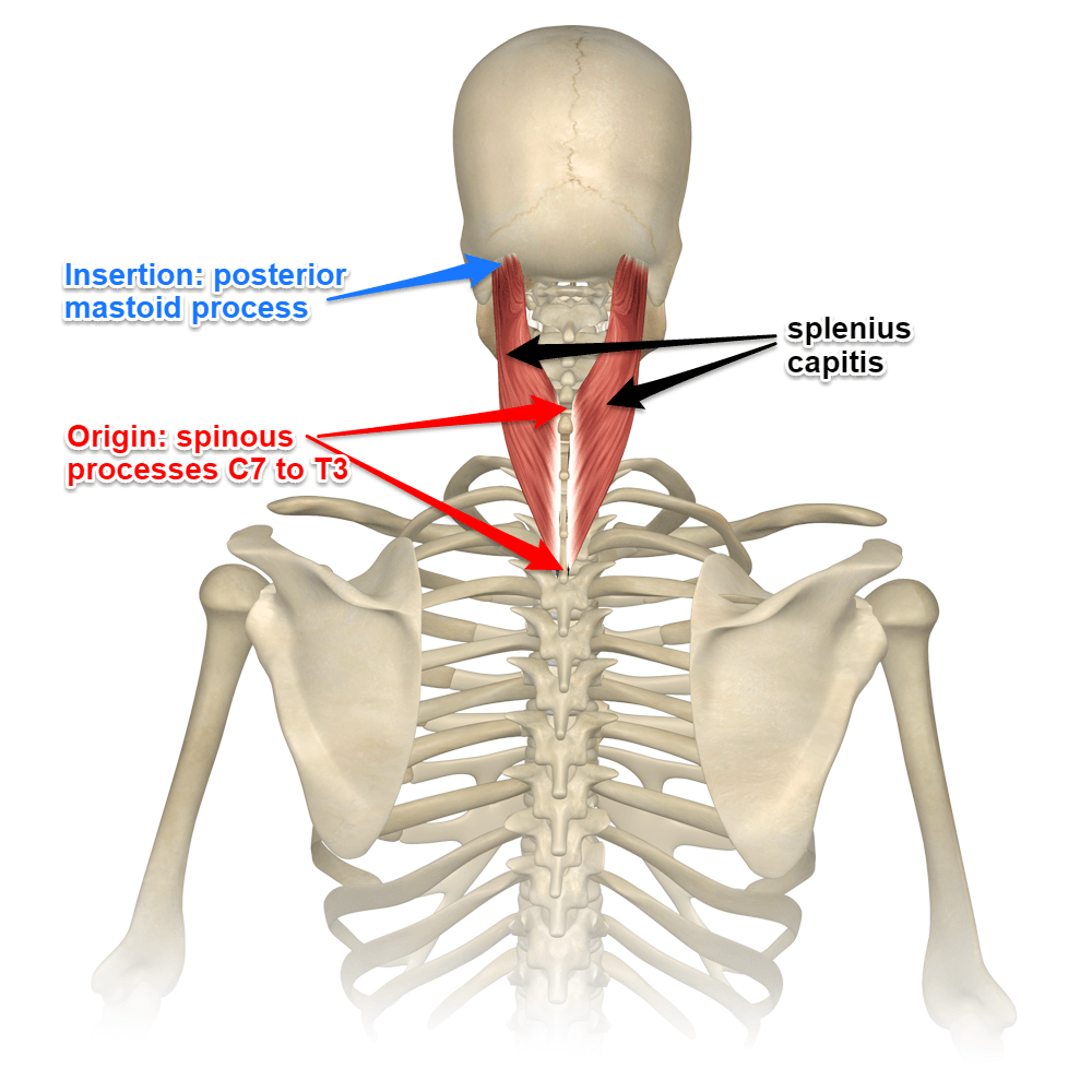
There are several individual muscles within the back anatomy, and it's important to take a quick look the image below to shows all the major back muscles (as well as some neck muscles)
The three scalene muscles are found forming the floor of the posterior triangle. William is a final year medical student in australia who has taught anatomy to tertiary science and medical students since 2010. They start at the top of the neck and go down to the tailbone. Only two of the more obvious and superficial neck muscles are. Cervical spine anatomy is quite complex. Some neck muscles attach to the clavicles. Muscles of the shoulder and back laminated anatomy chart. Together they extend neck, and individually they draw and rotate head to one side i.e. The major muscle of the back of the neck, the trapezius, is involved in movements of the scapula and is dealt with in the next section, on the muscles in this view of a male figure with one arm up and one arm on the hip, there is a tremendous number of clearly defined anatomical shapes, large and small. The superficial group acts on upper limbs and. Several other muscles of the back also extend up to the neck region and are partly connected with the cervical part of the vertebral column, including the trapezius, levator scapulae, splenius, iliocostalis, longissimus, rotatores, semispinalis, interspinales, and intertransversarii muscles. Sternohyoid, sternothyroid, thyrohyoid, omohyoid anterior vertebral muscles: Neck mobility is necessary primarily to rotate the head and keep the head upright.
12 photos of the anatomy of neck muscles back of neck anatomy. There are around 650 skeletal muscles within the typical human body.


0 Komentar